
Diseases & Treatments, Health & General Care, Veterinary Science & Advances
Pet Imaging and Its Vital Role in Diagnosing Diseases
Jun
In modern veterinary medicine, pet imaging is undeniably one of the most effective diagnostic tools available. In fact, it provides critical insights when clinical signs are vague or nonexistent. Pets are unable to describe their discomfort or symptoms, which makes accurate diagnosis challenging. Therefore, imaging allows veterinarians to examine the animal’s internal systems without invasive methods. From evaluating bones and joints to diagnosing internal organ issues, nervous system disorders, and tumor growth, these imaging methods are crucial. Techniques such as X-rays, ultrasound, CT scans, and MRI not only improve diagnostic precision but also do so in a non-invasive and timely manner. As a result, pet imaging has become essential in delivering fast and effective treatment in modern animal healthcare.

The Importance of Pet Imaging in Early Disease Detection
Pet imaging plays a critical role in detecting diseases early—when they are still treatable. These methods help veterinarians identify internal, skeletal, or neurological problems even before symptoms become severe. In emergency situations such as trauma, sudden pain, or loss of consciousness, imaging can save valuable time and prevent complications. Moreover, in pets without visible symptoms, imaging is often the only tool that reveals what’s going wrong internally. As a result, timely use of pet imaging improves diagnosis, supports better treatment decisions, and speeds up recovery.
Types of Pet Imaging Techniques:
- X-ray (Radiography)
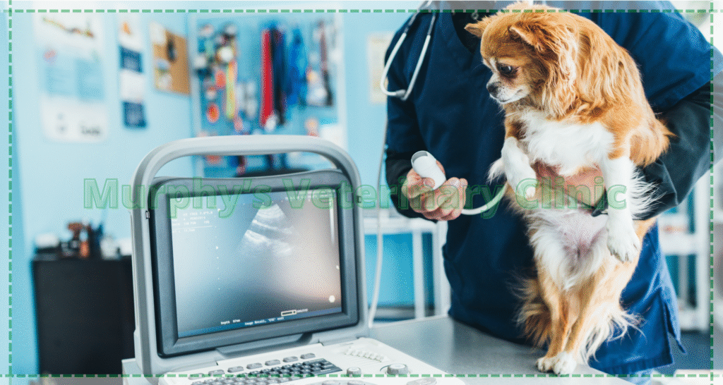
- Ultrasound
- CT-Scan
- MRI
- Echocardiography
- Dental X-ray
Applications of Pet Imaging in Evaluating Organs and Tissues
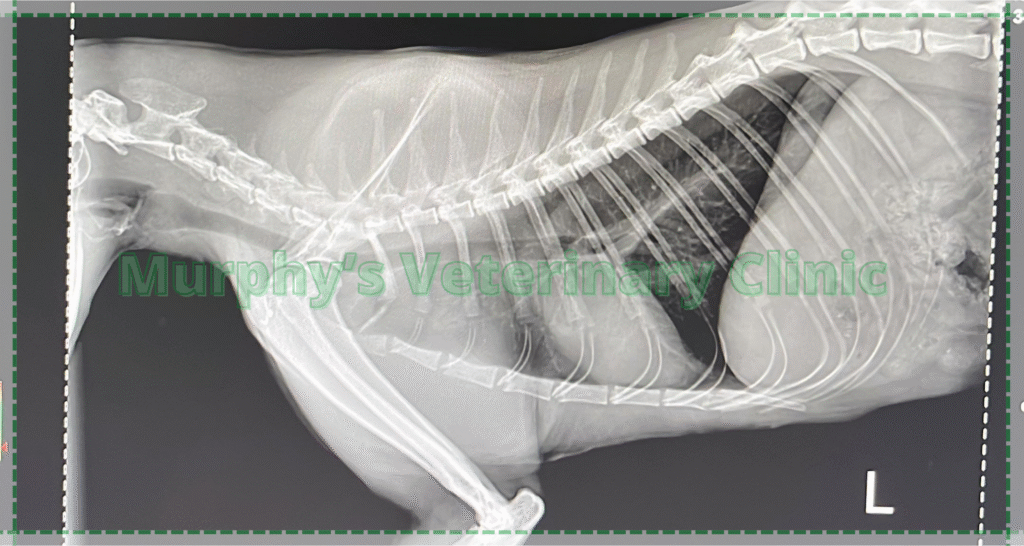
Pet imaging allows veterinarians to assess internal organs and tissues thoroughly and without surgical intervention. For example, when examining soft organs like the liver, kidneys, bladder, and stomach, ultrasound is often the best choice due to its safety and real-time results. CT or MRI may be required for deeper tissue investigation or precise evaluation of growths. Meanwhile, X-ray imaging remains useful for visualizing the heart and lungs, especially in animals experiencing respiratory symptoms. Therefore, the use of appropriate imaging methods helps ensure a more accurate diagnosis and targeted treatment.
Here are some examples of applications of imaging of pets:
Digestive System Evaluation
Imaging can detect blockages, inflammation, or masses in the intestines and stomach.
Liver and Kidney Assessment
Structural or functional changes in these organs can be identified with high precision.
Respiratory System Examination
The lungs, trachea, and chest cavity can be examined non-invasively.
Tumor Growth Monitoring
If a mass is found, its size, spread, and progression can be closely tracked.
Types of Pet Imaging and Their Features
Each imaging method, therefore, serves a specific purpose depending on the condition and the body part being evaluated. The following table compares the most commonly used pet imaging techniques based on their application, benefits, and limitations.
| Imaging Type | Main Application | Advantages | Limitations |
| X-ray | Bones, lungs, teeth | Fast, affordable | Limited for soft tissue details |
| Ultrasound | Soft organs (liver, bladder, etc.) | Radiation-free, safe in pregnancy | Quality depends on technician skill |
| CT Scan | Tumors, complex fractures | High detail, 3D visualization | Expensive, requires anesthesia |
| MRI | Brain and spinal cord | Best for neurological structures | Expensive, needs special equipment |
Role of Pet Imaging in Diagnosing Skeletal and Movement Disorders
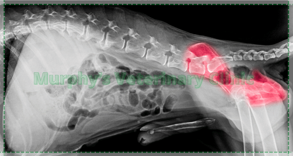
Pet imaging is highly valuable in detecting skeletal and musculoskeletal conditions. For example, X-rays can quickly identify bone fractures, dislocations, or deformities. In more complex situations, CT scans provide detailed 3D imaging of the affected area. Consequently, this helps veterinarians plan appropriate surgical interventions, physical therapy routines, or immobilization strategies to ensure proper recovery.
Fracture Detection
Clearly shows the location and severity of broken bones.
Joint and Arthritis Analysis
Identifies changes in joint spacing and surrounding bone structures.
Dislocation and Misalignment
Helps spot bone or joint displacement without exploratory surgery.
Post-Surgery Evaluation
Monitors healing and placement of screws, plates, or bone fusions.
How Pet Imaging Helps in Diagnosing Neurological and Brain Disorders
When pets show signs of seizures, disorientation, difficulty walking, or sudden behavior changes, pet imaging becomes essential. As a result, advanced imaging like MRI or CT is used to investigate the brain and spinal cord for tumors, injuries, or abnormalities. These scans offer deep insights that cannot be gained from physical examination alone, enabling early and effective intervention.
| Condition | Common Symptoms | Best Imaging Method | Imaging Benefits |
| Sudden Seizures | Tremors, loss of consciousness | MRI | Detects tumors, inflammation, or brain injury |
| Paralysis or Weak Limbs | Immobility, numbness | MRI or CT | Identifies spinal cord or brain damage |
| Severe Behavioral Issues | Aggression, confusion, fear | MRI | Reveals brain structure and hidden conditions |
| Congenital Abnormalities | Birth defects, skull deformities | MRI or CT | Diagnoses inherited or structural nerve issues |
Conclusion
Using pet imaging is one of the most advanced and necessary diagnostic tools in modern veterinary care. It allows veterinarians to detect internal problems without invasive methods. In many cases, pet imaging helps save an animal’s life and guides precise treatment plans. From simple fracture checks to complex neurological assessments, imaging plays a vital role. By choosing the right technique, diagnosis becomes faster and more accurate, leading to more effective and timely treatment. Pet owners should understand the value of imaging and seek it promptly when any abnormal signs appear.
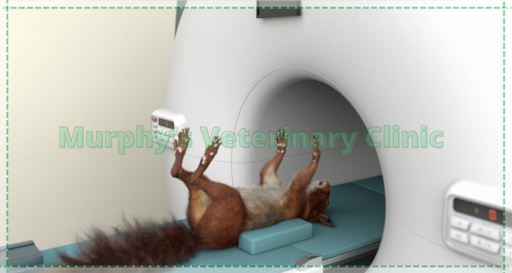
Murphy’s Veterinary Clinic is equipped with advanced pet imaging tools like digital ultrasound and digital radiography to provide fast and accurate diagnoses. Our experienced team of veterinarians interprets each image with precision to guide the most effective treatment. Whether your pet needs internal evaluation, bone assessment, or neurological imaging, Murphy’s is here to deliver expert care with compassion.
